Parasitism and its Effects on Equine Gastrointestinal Health
Parasitic challenge in horses remains a significant threat to health and performance. Over the years, various regular de-worming programs have failed to eliminate this problem and now there is the omnipresent issue of increasing anthelmintic resistance. How we manage the parasitic burden is again being scrutinized and, in the light of the new AAEP Parasite Control Guidelines, recommendations are to use rigorous testing and monitoring to address the challenge. One of the most concerning aspects of parasitic burden is the damage caused to the gastrointestinal (GI) tract, a likely underestimated issue. In line with the new guidelines, the need for ongoing monitoring, as well as early detection technologies is great.This article describes the various types of parasites that can affect the equine GI tract and resulting pathologies and discusses modern approaches to controlling the parasitic challenge.
Types of parasites affecting the GIT
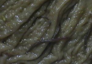
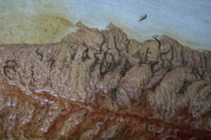
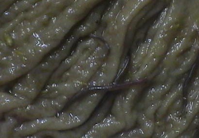
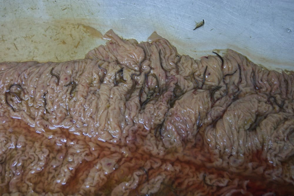
The main groups of parasites affecting equids are the large strongyles, small strongyles, ascarids and tapeworms (Nielsen et al., 2014). However, protozoan species are also implicated. Several of the major parasites have significant prepatent periods (2-12 months depending on species) (Andersen et al., 2013).Strongyles in Colonic MucosaLarge strongyles, known also as blood or red worms, enter the horse via ingestion of infective larvae from feces. After emerging in the small intestine they consequently migrate through body tissues before returning to the large intestine to mature. This can cause significant damage with larvae found in the liver, and in perirenal, peritoneal and pancreatic tissues. Adult strongyles are blood feeders and can cause extensive damage to the intestine wall that can result in anemia, as well as weight loss and diarrhea (Merck Veterinary Manual, 2010).Small strongyles, or cyathostomins, differ from their larger counterparts in that they remain within the intestinal tract. They develop in the gut wall and emerge as stage L4 larvae. This emergence may cause slight damage to the intestinal wall but cyathostomins are less severe in their effects than the blood worms (Merck Veterinary Manual, 2010). However, if there is mass emergence of these larvae then significant damage can ensue. This situation is known as larval cyathostominosis (Nielsen, 2012; Andersen et al., 2013). Co-infection of large and small strongyles is common.Small Strongyles in Right Dorsal ColonAscarids mainly infect young animals via ingestion from contaminated pasture, the most notable of which is Parascaris equorum (large roundworm). This parasite migrates through the liver and lungs before returning to the small intestine (Andersen et al., 2013). Ill-thrift, lethargy with the potential for impaction colic are often signs of infection. In severe challenges, respiratory signs can also be noted (Andersen et al., 2013).Tapeworms (cestodes) are again found in the intestines, both small and large. Often there are no clinical signs with mild infections but anemia, weight loss, ulceration and digestive aberrations can result from more severe challenges. Indeed, colic appears more likely in those animals with tapeworm infestation and they can easily be seen at necropsy at the ileocecal junction (Andersen et al., 2013).
Symptoms
Mild symptoms for most parasitic infections involve weight and condition loss, ill-thrift, lethargy and weakness. More severe infections can result in:
anemia from blood loss, for example with adult large strongyles
gastric and colonic ulceration from damage to the mucosal lining
enteritis
colic
thrombosis
respiratory distress
problems associated with damage to tissues due to migration of the larvae.
Many parasitic pathologies are associated with the gastrointestinal tract and, therefore, pose a significant threat to the horse’s well-being even without clinical signs. Gastric ulcers have received significant attention over the last few years with numerous cited causes, parasitic infection being one. Bot eggs and their subsequent larvae can cause substantial damage to the mucosal lining resulting in ulceration.Less well-known is the incidence of colonic ulcers (Pellegrini, 2005). Again, damage to the protective mucosal lining of the colon, for example cyathostomins that develop in the gut wall, can lead to the formation of intestinal ulcers.Of a more serious nature, Strongyles vulgaris migrates to the mesenteric artery and can significantly restrict blood flow to the digestive tract leading to reduced digestive function, tissue necrosis and, ultimately, colic. Intestinal impaction and sometimes perforation have also been known with heavy parasitic infections.Any damage, not matter how mild, to the digestive tract compromises digestive function and the well-being of the horse. While heavy and severe parasitic infections are more easily spotted, mild issues can create gastrointestinal aberrations without necessarily showing definitive clinical signs. Reduced nutrient absorption, dysbiosis in the hind gut and a reduced ability to deal with other intestinal pathogens can all result from compromised digestive function.
New AAEP guidelines for parasite control
The Association of American Equine Practitioners (AAEP) have recently proposed a set of guidelines for the control of parasites in horses. They highlight that much has changed in the parasitic fauna of the horse since existing strategies for control were devised and there is now widespread resistance to anthelmintics in small strongyles and the ascarid, Parascaris equorum. Therefore, current recommendations are based on selective therapy followed by monitoring and periodic re-testing:
A faecal egg count reduction test (FECRT) is performed to assess levels of resistance. Egg counts are performed prior to treatment with an anthelmintic and the test repeated 14 days following treatment.
Level of resistance is then calculated using the level of reduction in the egg counts.
Subsequently, the egg reappearance period (ERP) (time between last effective anthelmintic treatment and reappearance of significant egg shedding) is monitored using periodic re-testing.
Early detection of issues
Parasitic burden is usually estimated by carrying out an FEC and then using further methods to identify individual species. Treatment with appropriate anthelmintics is then usually indicated where the burden is high enough. However, there is limited evidence for the current threshold values for number of eggs and these thresholds were decided without any real knowledge of how the egg count related to the challenge in the animal (Neilsen et al., 2014). In a study looking at strongyle infection post-mortem, Nielsen et al. (2010) noted no direct linear relationship between the FEC results and actual burden in the horse. In fact, some horses with seemingly low egg counts were found to have high worm burdens inside. As a result, FEC cannot be used as a direct indication of the requirement for anthelmintic treatment (Nielsen et al., 2014). Additionally, some parasites, such as pinworms, do not shed eggs in the feces yet are a common worm found in horses. Thus, this selective therapy approach is designed to help reduce development of resistance but requires vigilant surveillance and monitoring (Nielsen, 2012), which is reflected in the new AAEP guidelines.That said, damage of, and, therefore, compromise to, the intestine can occur even with low FEC results and early detection of gastrointestinal aberrations should be encouraged to minimize negative effects. One such study investigated the potential relationship between fecally excreted albumin and parasitic burden using a commercially available lateral flow immunoassay containing monoclonal antibodies targeted to equine hemoglobin and albumin (SUCCEED™ FBT™, Freedom Health, Ohio, USA) (Kerbyson et al., 2014). The study outline was as follows:
The FBT test was carried out prior to administration of appropriate anthelmintic after establishment of parasitic burden.
Then a subsequent test was carried out on the same horses 14 days post-anthelmintic administration.
More fecal albumin was detected before anthelmintic treatment compared with after treatment but was not associated with actual degree of parasite burden. This study is currently being conducted on a larger scale, however, it demonstrates the potential for early detection of gastrointestinal damage from parasites. Obviously, albumin in feces is not only generated by parasites so presence of albumin indicates the need for further investigation with parasites being one area to look at.
Support intestinal health while treating for parasitism
While anthelmintics will treat the parasitic challenge, they will not aid the digestive tract to recover. Given that varying levels of damage to the digestive tract can occur, it would be prudent in this situation to ensure overall intestinal function is supported throughout treatment and beyond. Clearly, the basic principles of feeding should apply but promoting a healthy absorptive surface, as well as a stable hindgut environment should be key targets for intestinal support.
Conclusion
Internal parasites have always been an issue in horses with the main categories being large and small strongyles, ascarids and tapeworms.
Symptoms can range from weight loss and lethargy to tissue damage and colic.
Gastrointestinal damage is common with parasitic burdens, including gastric and colonic ulcers and damage to the gut wall.
AAEP guidelines suggest selective therapy with continual monitoring.
Use of a field test, such as an FBT, can aid in early detection of parasitic burdens
Digestive health should be supported throughout and following treatment for parasites.
References
Andersen, U.V. et al. (2013) Recent advances in diagnosing pathogenic equine gastrointestinal helminths: The challenge of prepatent detection Veterinary Parasitology 192: 1-9Kerbyson, N. et al. (2014) The effect of parasite burden on faecally excreted albumin in horses ACVIM ProceedingsThe Merck Veterinary Manual (2010) Tenth edition. C.M. Kahn (Ed). Merck and Co., Inc. Whitehouse Station, NJ, USANielsen, M.K. et al. (2010) Analysis of multiyear studies in horses in Kentucky to ascertain whether counts of eggs and larvae per gram of feces are reliable indicators of numbers of stronglyes and ascarids present Veterinary Parasitology 174: 77-84Nielsen, M.K. (2012) Sustainable equine parasite control: Perspectives and research needs Veterinary Parasitology 185: 32-44Nielsen, M.K. et al. (2014) Selective therapy in equine parasite control – Application and limitations Veterinary Parasitology 202: 95-103Pellegrini, F.L. (2005) results of a large-scale necroscopic study of equine colonic ulcers Journal of Equine Veterinary Science 25(3): 113-117